Abdominal aortic aneurysm
Permanent focal dilatation of the abdominal aorta (≥3 cm) from medial wall weakening
Overview
A focal dilatation of the abdominal aorta to ≥3 cm, usually infrarenal and degenerative. Most are asymptomatic and found on screening (the NHS offers a one-off ultrasound to men at 65); the catastrophe is rupture. Elective repair is offered above a size/growth threshold, balancing rupture risk against operative risk.
Recognise
- Usually asymptomatic; a pulsatile, expansile central abdominal mass; back/abdominal pain if symptomatic/expanding
- Ruptured AAA — the classic triad: sudden severe abdominal/back pain, hypotension/collapse, and a pulsatile mass
- Risk factors: male sex, age, smoking, hypertension, family history, atherosclerosis
Red flags
- Suspected rupture (pain + collapse + pulsatile mass) → immediate vascular surgery; do NOT delay a haemodynamically unstable patient for a CT
- Tender or rapidly expanding aneurysm → symptomatic AAA → urgent repair
Differentials & how to tell them apart
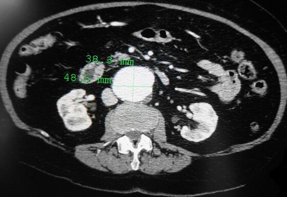
Abdominal aortic aneurysm — dilated aorta on contrast CT
James Heilman, MD / CC BY-SA 3.0 — Wikimedia Commons
Investigations
Ultrasound (screening and surveillance — the first-line test); CT angiography for surgical planning and suspected rupture (only if stable). Cardiovascular risk assessment before elective repair.
Management
Surveillance + risk factors for small AAA; elective repair (EVAR/open) at ≥5.5 cm or symptomatic
- 1Detect on screening/ultrasound; for small aneurysms enter surveillance and modify risk factors (smoking cessation, statin, BP control).Gate: Suspected rupture (sudden pain + hypotension + pulsatile mass) → straight to theatre/vascular surgery; an unstable patient should not be delayed for imaging.
- 2Refer for elective repair at ≥5.5 cm, if symptomatic/tender, or growing >1 cm/year — choosing EVAR or open repair based on anatomy and fitness.
Key points
Pulsatile expansile abdominal mass = AAA; screen men at 65 with ultrasound. Sudden abdominal/back pain + collapse + pulsatile mass = rupture → theatre, not CT, if unstable. Never accept 'renal colic' in an older arteriopath without excluding a ruptured AAA. Elective repair at ≥5.5 cm.
Monitor & prognosis
Ultrasound surveillance by size; cardiovascular risk optimisation; post-repair endoleak surveillance (EVAR).
Excellent if electively repaired; ruptured AAA has very high mortality.
Source: NICE NG156 (AAA); NHS AAA screening