Bone tumours & metastases
Primary bone malignancy (osteosarcoma, Ewing, chondrosarcoma) or — far commoner — metastasis/myeloma
Overview
Bone malignancy is far more often secondary (metastases from breast, prostate, lung, kidney, thyroid; and myeloma) than primary. Primary bone sarcomas — osteosarcoma (adolescents, around the knee), Ewing sarcoma (children/young, diaphysis), chondrosarcoma (older) — are rare but important. Persistent bone pain (especially night pain), a pathological fracture or a destructive lesion should prompt urgent investigation.
Recognise
- Persistent localised bone pain, often worse at night and not relieved by rest; a mass; pathological fracture
- Osteosarcoma: adolescent, metaphysis around the knee, 'sunburst'/Codman triangle on X-ray; Ewing: diaphysis, 'onion-skin' periosteal reaction, systemic features
- Metastases: known primary (breast/prostate/lung/kidney/thyroid), often multiple; myeloma → lytic lesions + hypercalcaemia + renal impairment + anaemia
Red flags
- Metastatic spinal cord compression (back pain + neurology) → emergency MRI + dexamethasone + oncology (cross-ref acute_care/neurology)
- Hypercalcaemia of malignancy (confusion, polyuria, dehydration) → emergency
- Pathological fracture / lytic lesion → urgent investigation and orthopaedic input
Differentials & how to tell them apart
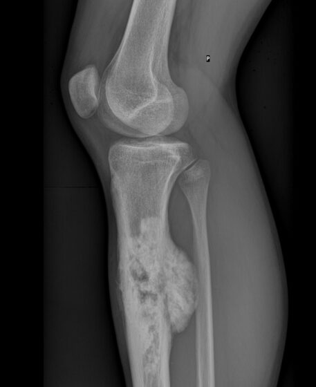
Osteosarcoma of the tibia — aggressive periosteal reaction (X-ray)
Yousef Samir / CC BY-SA 4.0 — Wikimedia Commons
Investigations
X-ray (lytic/sclerotic lesion, periosteal reaction); MRI (local staging) and CT/bone scan/PET (staging); biopsy at a specialist sarcoma centre (do NOT biopsy through compartments that compromise later surgery); myeloma screen (serum/urine electrophoresis, calcium, renal function); find the primary for metastases.
Management
Urgent imaging + specialist MDT; treat metastases/myeloma per primary; emergency care for cord compression/hypercalcaemia
- 1Investigate persistent/night bone pain or a destructive lesion with X-ray then MRI/staging; biopsy at a specialist centre (avoid compromising future surgery). Screen for myeloma and identify the primary in suspected metastases.Gate: Metastatic spinal cord compression (back pain + neurology) or hypercalcaemia of malignancy → emergencies needing immediate dexamethasone/MRI/oncology — do not delay.
- 2Primary sarcomas → neoadjuvant chemotherapy + limb-sparing surgery via MDT; metastatic disease → treat the primary, bisphosphonates/denosumab, radiotherapy for pain, and orthopaedic stabilisation of fractures.
Key points
Persistent bone pain, worse at night, ± pathological fracture = bone tumour until excluded. Far more often metastasis/myeloma than primary sarcoma. Osteosarcoma (teen, knee, sunburst); Ewing (diaphysis, onion-skin). Emergencies: cord compression (dexamethasone + MRI) and hypercalcaemia. Biopsy only at a sarcoma centre.
Monitor & prognosis
Staging response, skeletal events, calcium; MDT follow-up.
Primary sarcomas: improved with modern chemo + surgery; widespread metastatic disease often palliative.
Source: NICE NG12 (suspected cancer); sarcoma MDT; cross-ref endocrine (hypercalcaemia)