Fractures & pathological fractures
Break in bone cortex — traumatic, fragility (osteoporotic), or pathological (through abnormal bone)
Overview
A breach of bone continuity. Most follow trauma; a fragility fracture follows low-energy trauma (osteoporosis); a pathological fracture occurs through abnormal bone (metastasis, myeloma, primary tumour, Paget's) with minimal or no trauma. Management follows reduce–hold–rehabilitate, with attention to the soft tissues and the underlying bone health or pathology.
Recognise
- Pain, swelling, deformity, loss of function; localised tenderness, abnormal movement/crepitus
- X-ray confirms: describe site, pattern (transverse/oblique/spiral/comminuted), displacement, angulation, intra-articular involvement, open vs closed
- Pathological fracture: minimal trauma, prior bone pain, known cancer, lytic lesion on X-ray
Red flags
- Open fracture (wound communicating with bone) → IV antibiotics, tetanus, photograph + cover, urgent orthoplastics
- Neurovascular compromise distal to the fracture, or compartment syndrome → emergency
- Pathological fracture → investigate the underlying cause (metastasis/myeloma)
Differentials & how to tell them apart
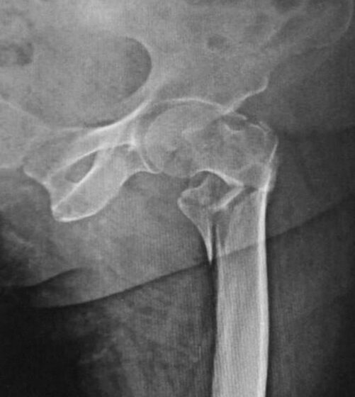
Comminuted hip (neck-of-femur) fracture (X-ray)
R. R. Memon et al. / CC BY 4.0 — Wikimedia Commons
Investigations
X-ray (two views, joint above and below); CT for complex/intra-articular fractures; MRI/bone scan if pathological/occult (e.g. scaphoid, hip); assess neurovascular status; bloods + myeloma/metastasis work-up for pathological fractures; DEXA for fragility fractures.
Management
Reduce + immobilise + analgesia; open fracture → antibiotics + debridement; treat the underlying bone
- 1Assess neurovascular status and the soft tissues; confirm and characterise on X-ray. Provide analgesia, reduce and immobilise. Open fractures → IV antibiotics, tetanus and urgent orthoplastic management.Gate: Neurovascular compromise or compartment syndrome distal to the fracture is an emergency; a pathological fracture (minimal trauma/lytic lesion) needs investigation of the underlying cause before fixation.
- 2Definitive fixation for unstable/displaced/intra-articular fractures, then rehabilitation; treat the underlying bone (osteoporosis after a fragility fracture, oncology for pathological fractures).
Key points
Reduce–hold–rehabilitate. Always check and document neurovascular status and the soft tissues. Open fracture → antibiotics + tetanus + debridement. A fragility fracture mandates osteoporosis treatment; a pathological fracture (minimal trauma + lytic lesion) mandates a malignancy/myeloma work-up.
Monitor & prognosis
Healing on serial X-ray (union), neurovascular status, complications, bone-health treatment.
Most heal well; complications (non-union, malunion, AVN, infection) and underlying pathology modify outcome.
Source: BOAST/NICE NG38; cross-ref osteoporosis & bone tumours