Central retinal artery occlusion
Embolic/thrombotic occlusion of the central retinal artery (a retinal 'stroke')
Overview
Sudden, painless, profound monocular visual loss from occlusion of the central retinal artery — effectively a retinal stroke. Causes: carotid/cardiac embolism, and crucially giant cell arteritis. Fundus shows a pale retina with a cherry-red spot and an RAPD. Time-critical, with a poor prognosis; treat as a stroke/TIA equivalent and exclude GCA.
Recognise
- Sudden, painless, severe LOSS of vision in one eye (often counting-fingers or worse)
- Relative afferent pupillary defect (RAPD); fundus: pale oedematous retina with a CHERRY-RED SPOT at the macula; segmented 'cattle-trucking' in vessels
- Amaurosis fugax (transient curtain) may precede it; embolic source (carotid, AF) or GCA
Red flags
- CRAO is a stroke equivalent → urgent stroke pathway (carotid imaging, ECG, vascular risk) AND exclude GCA (ESR/CRP) in the older patient — bilateral blindness risk
- Jaw claudication, scalp tenderness, headache → arteritic cause: high-dose steroids immediately
Differentials & how to tell them apart
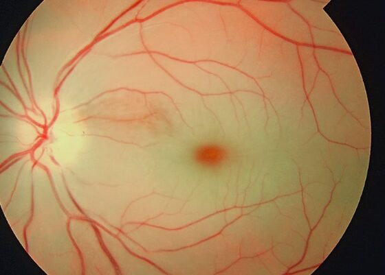
CRAO — pale retina with a cherry-red spot at the macula
Dr. Gopal Bisht / CC BY-SA 4.0 — Wikimedia Commons
Investigations
Urgent ESR/CRP (exclude GCA), fundoscopy, carotid Doppler, ECG/echo for embolic source; same as TIA work-up.
Management
Emergency referral; exclude GCA (steroids if arteritic) + stroke-pathway work-up
- 1Emergency ophthalmology/stroke assessment. Acute ocular-massage / IOP-lowering (acetazolamide, anterior-chamber paracentesis) are sometimes attempted within hours but evidence is limited.Gate: In anyone ≥50, CHECK ESR/CRP and treat as GCA with high-dose steroids if suspected BEFORE the fellow eye is lost; otherwise manage as a TIA/stroke equivalent (carotid imaging, ECG, vascular prevention)
- 2Vascular secondary prevention (antiplatelet, statin, BP/AF management); carotid endarterectomy for significant ipsilateral stenosis.
Key points
Sudden painless monocular blindness + RAPD + cherry-red spot = CRAO. It is a stroke — work it up as one, and never miss GCA in the older patient.
Monitor & prognosis
Vascular risk-factor management; GCA follow-up if arteritic.
Poor visual recovery; the priority is the brain and the fellow eye.
Source: RCOphth; cross-ref neurology (stroke/GCA)