Central retinal vein occlusion
Thrombosis of the central retinal vein (hypertension, hyperviscosity, glaucoma)
Overview
Occlusion of the central retinal vein causing sudden, painless, variable monocular visual loss. The fundus shows widespread flame and dot-blot haemorrhages, dilated tortuous veins, cotton-wool spots and disc/macular oedema ('blood and thunder' / stormy-sunset fundus). Risk factors: hypertension, diabetes, hyperviscosity, glaucoma. Treated with anti-VEGF for macular oedema; watch for neovascular complications.
Recognise
- Sudden painless blurring/loss of vision in one eye, usually less severe than CRAO and variable
- Fundus: widespread retinal HAEMORRHAGES in all four quadrants, dilated tortuous veins, cotton-wool spots, optic-disc and macular oedema
- Ischaemic CRVO (poor vision, RAPD, many cotton-wool spots) risks rubeosis → neovascular glaucoma
Red flags
- Ischaemic CRVO → neovascularisation of the iris (rubeosis) and neovascular glaucoma at ~3 months ('100-day glaucoma') — needs panretinal photocoagulation
- Bilateral CRVO/young patient → investigate hyperviscosity/thrombophilia
Differentials & how to tell them apart
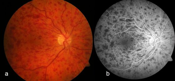
CRVO — widespread 'blood and thunder' retinal haemorrhages
Werner JU, Böhm F, Lang GE / CC BY 4.0 — Wikimedia Commons
Investigations
Fundoscopy; OCT (macular oedema); fluorescein angiography (ischaemic vs non-ischaemic). BP, glucose/HbA1c, lipids, IOP; hyperviscosity screen if young/bilateral.
Management
Refer; anti-VEGF for macular oedema + treat vascular risk factors
- 1Refer to ophthalmology. Treat the macular oedema (intravitreal anti-VEGF first-line, or steroid implant) and manage vascular risk factors (BP, diabetes, lipids, IOP).Gate: Identify ISCHAEMIC CRVO (poor vision, RAPD, extensive non-perfusion) — it risks rubeosis and neovascular glaucoma, requiring monitoring ± panretinal photocoagulation
- 2Neovascular complications → panretinal photocoagulation; lifelong vascular risk management; investigate young/bilateral cases for thrombophilia/hyperviscosity.
Key points
Sudden painless monocular blur + 'blood and thunder' four-quadrant haemorrhages = CRVO. The danger downstream is neovascular glaucoma — distinguish ischaemic from non-ischaemic.
Monitor & prognosis
Serial OCT/visual acuity; monitor for neovascularisation.
Variable; non-ischaemic better than ischaemic.
Source: RCOphth Retinal vein occlusion guidelines