Keratoconus
Progressive corneal thinning and conical ectasia (often atopic; eye-rubbing)
Overview
Progressive non-inflammatory thinning and conical protrusion of the cornea, presenting in teens/twenties with worsening irregular (myopic) astigmatism. Associated with atopy/eye-rubbing, Down and connective-tissue syndromes. Managed with rigid/scleral lenses and corneal cross-linking to halt progression.
Recognise
- Progressive blurring with frequent spectacle changes and irregular astigmatism, often bilateral/asymmetric
- Signs: corneal thinning, Vogt striae, Fleischer ring, Munson sign (lower-lid bulge on downgaze), scissoring retinoscopy reflex
- Acute hydrops: sudden painful corneal oedema from Descemet rupture
Red flags
- Acute hydrops (sudden pain, marked oedema and visual loss) → ophthalmology
Differentials & how to tell them apart
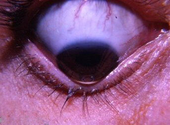
Keratoconus — conical cornea (Munson sign on downgaze)
William Charles Caccamis / CC BY-SA 4.0 — Wikimedia Commons
Investigations
Corneal topography/tomography (the diagnostic test — shows inferior steepening/thinning); refraction.
Management
Rigid/scleral contact lenses; corneal cross-linking to halt progression
- 1Spectacles/soft lenses early; rigid gas-permeable or scleral lenses for irregular astigmatism. Stop eye-rubbing.Gate: Document progression on topography — progressive disease is offered corneal collagen cross-linking to halt it before vision is lost
- 2Corneal cross-linking for progression; intacs; corneal transplant (deep anterior lamellar or penetrating keratoplasty) for advanced disease/scarring.
Key points
Young patient + rapidly changing astigmatism + atopy/eye-rubbing = keratoconus. Topography makes the diagnosis; cross-linking stops it progressing.
Monitor & prognosis
Serial topography for progression.
Often stabilises; cross-linking improves the course.
Source: College of Optometrists CMG; RCOphth