Papilloedema
Bilateral optic-disc swelling from raised intracranial pressure
Overview
Optic-disc swelling caused by raised intracranial pressure — by definition BILATERAL. It is a sign, not a diagnosis: the task is to find and treat the cause (space-occupying lesion, idiopathic intracranial hypertension, venous sinus thrombosis, meningitis, malignant hypertension). Vision is preserved early but chronic papilloedema causes progressive field loss.
Recognise
- Bilateral optic-disc swelling: blurred disc margins, elevated disc, loss of spontaneous venous pulsation, peripapillary haemorrhages
- Headache (worse on waking/lying/Valsalva), nausea/vomiting, transient visual obscurations, pulsatile tinnitus, diplopia (VI palsy = false-localising sign)
- Enlarged blind spot; central acuity preserved until late
Red flags
- Papilloedema + focal neurology → urgent neuroimaging for a space-occupying lesion/haemorrhage
- IIH with progressive field loss → urgent treatment to save vision; consider CVST
Differentials & how to tell them apart
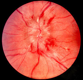
Papilloedema — optic-disc swelling with blurred margins
Jonathan Trobe, M.D. / CC BY 3.0 — Wikimedia Commons
Investigations
Urgent neuroimaging (MRI/CT + venography to exclude mass/CVST) FIRST, then lumbar puncture for opening pressure (raised) and contents (only after imaging excludes a mass). Visual fields; BP.
Management
Urgent neuroimaging to find the cause; treat the cause (e.g. acetazolamide for IIH)
- 1Treat as raised ICP: urgent neuroimaging (MRI + venography) to exclude a mass or venous sinus thrombosis.Gate: Do NOT perform lumbar puncture until imaging has EXCLUDED a space-occupying lesion (coning risk); LP then confirms raised opening pressure in IIH
- 2Cause-specific: IIH → weight loss + acetazolamide (± shunt/optic-nerve-sheath fenestration if vision threatened); tumour → neurosurgery; CVST → anticoagulation; monitor fields to protect sight.
Key points
Bilateral disc swelling = raised ICP until proven otherwise. Image before you LP. Central vision is preserved early — an enlarged blind spot and field loss are the warning signs.
Monitor & prognosis
Serial visual fields and disc appearance; treat the cause.
Vision preserved if pressure controlled; chronic papilloedema causes atrophy.
Source: RCOphth; NICE (IIH); neurology guidance