Hydronephrosis & obstructive uropathy
Obstruction to urine flow → back-pressure dilatation of the collecting system ± renal impairment
Overview
Dilatation of the renal pelvis and calyces (hydronephrosis) from obstruction to urine flow, which may be upper-tract (stone, PUJ obstruction, tumour, retroperitoneal fibrosis) or lower-tract (prostate, urethral stricture, pelvic malignancy, blocked catheter). The priorities are relieving the obstruction, watching for post-obstructive diuresis, and not missing an obstructed infected system.
Recognise
- Loin pain/fullness (upper-tract) or lower urinary symptoms/painful or painless retention (lower-tract); a palpable bladder in chronic retention
- AKI/CKD if bilateral or affecting a single kidney; anuria suggests complete bilateral or single-kidney obstruction
- Causes: stones, PUJ obstruction, ureteric/pelvic tumour, retroperitoneal fibrosis (upper); BPH/prostate cancer, urethral stricture, blocked catheter, neurogenic bladder (lower)
Red flags
- Obstructed + infected system → emergency decompression + antibiotics
- Post-obstructive diuresis after relieving chronic retention → monitor fluid/electrolytes and replace losses; high-pressure chronic retention with AKI → urgent catheter
Differentials & how to tell them apart
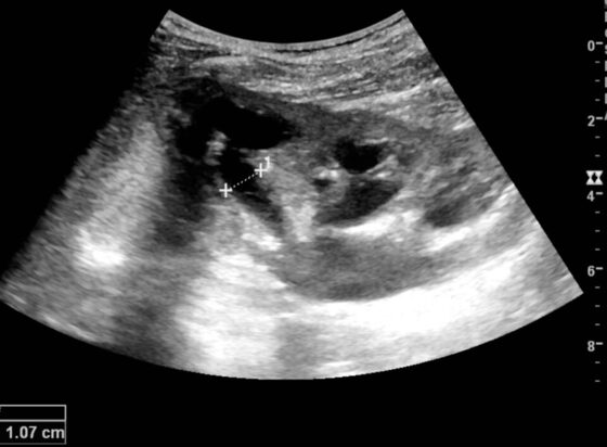
Hydronephrosis — dilated anechoic renal pelvis and calyces on ultrasound
Kristoffer Lindskov Hansen et al. / CC BY 4.0 — Wikimedia Commons
Investigations
Ultrasound (hydronephrosis, bladder volume — first-line); bladder scan (retention); U&Es (renal function); CT urogram to define the level/cause; urine dip/culture (infection); PSA/prostate exam or pelvic imaging for the cause.
Management
Relieve the obstruction (catheter / stent / nephrostomy) + treat the cause; watch post-obstructive diuresis
- 1Localise the level with a bladder scan and ultrasound (upper vs lower tract) and check renal function. Relieve the obstruction — catheter for lower-tract retention, nephrostomy/stent for upper-tract.Gate: An obstructed, infected system → emergency decompression + antibiotics; after relieving chronic retention, watch for post-obstructive DIURESIS and replace fluid/electrolyte losses.
- 2Treat the underlying cause (stone, prostate, tumour, stricture, retroperitoneal fibrosis) and manage any resulting CKD.
Key points
Hydronephrosis = obstruction until proven otherwise → bladder scan + USS to localise (upper vs lower), then relieve it (catheter / stent / nephrostomy) and treat the cause. Two traps: an obstructed INFECTED system (emergency decompression) and post-obstructive DIURESIS after relieving chronic retention (replace losses).
Monitor & prognosis
Renal function, urine output (post-obstructive diuresis), infection; resolution of hydronephrosis.
Recovery depends on duration/degree of obstruction; prolonged obstruction causes irreversible damage.
Source: NICE CKS; urology