Renal cancer
Renal cell carcinoma (clear-cell commonest) arising from the renal tubular epithelium
Overview
Renal cell carcinoma, usually clear-cell, arising from the proximal tubular epithelium. The classic (now uncommon) triad is haematuria, flank pain and a flank mass; most are found incidentally on imaging. It is notorious for paraneoplastic syndromes and for a left varicocoele (left renal vein/IVC involvement). Localised disease is treated surgically; it is relatively chemo/radio-resistant.
Recognise
- Classic triad (uncommon): visible haematuria + flank pain + palpable flank mass; often an incidental imaging finding; weight loss/fever
- Paraneoplastic: polycythaemia (EPO), hypercalcaemia (PTHrP), hypertension (renin), Stauffer syndrome (deranged LFTs without mets)
- A new LEFT-sided varicocoele (that doesn't empty when lying flat) → left renal vein obstruction by tumour; spreads to IVC/lung ('cannonball' metastases)
Red flags
- Visible haematuria → urgent referral (NICE NG12)
- Paraneoplastic hypercalcaemia/polycythaemia, IVC tumour thrombus, or a sudden left varicocoele → advanced disease work-up
Differentials & how to tell them apart
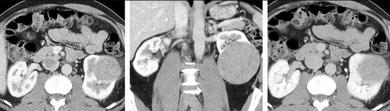
Renal cell carcinoma — enhancing renal mass on contrast CT
Guite & Hinshaw et al. / CC BY 3.0 — Wikimedia Commons
Investigations
CT abdomen/pelvis with contrast (the key diagnostic/staging test — enhancing renal mass); ultrasound (incidental detection); bloods — FBC (polycythaemia), calcium, LFTs, renal function; staging CT chest; biopsy in selected cases.
Management
Nephrectomy (partial/radical) for localised disease; TKI/immunotherapy for advanced
- 1Characterise a renal mass on contrast CT and stage (including CT chest). Visible haematuria → urgent referral. Localised disease → partial or radical nephrectomy.Gate: Recognise the paraneoplastic clues (polycythaemia, hypercalcaemia, hypertension) and a NEW LEFT VARICOCOELE (left renal vein obstruction) as pointers to RCC/advanced disease.
- 2Small/unfit → active surveillance or ablation; advanced/metastatic → tyrosine kinase inhibitors or immunotherapy (RCC is relatively chemo/radio-resistant); manage paraneoplastic features.
Key points
Haematuria + flank pain + flank mass (classic, uncommon) or an incidental enhancing renal mass = renal cell carcinoma → contrast CT + nephrectomy for localised disease. Think RCC with paraneoplastic polycythaemia/hypercalcaemia/hypertension and a NEW LEFT VARICOCOELE; 'cannonball' lung mets.
Monitor & prognosis
Post-nephrectomy surveillance, paraneoplastic markers, metastatic disease.
Good if localised and resected; relatively treatment-resistant when metastatic (TKI/immunotherapy improving).
Source: NICE NG12 (referral); renal cancer guidance