Urinary tract calculi
Stone formation in the renal tract (calcium oxalate commonest; also urate, struvite, cystine)
Overview
Stones in the renal tract causing renal/ureteric colic — sudden severe loin-to-groin pain with a restless patient unable to get comfortable. Most are calcium oxalate. Non-contrast CT KUB is the diagnostic gold standard. Management depends on stone size and site (NICE NG118), and the emergency is an obstructed, infected system requiring decompression.
Recognise
- Sudden severe colicky loin-to-groin pain, unable to lie still (vs peritonitis where they lie still), nausea/vomiting; visible or non-visible haematuria
- CT KUB: the stone and any hydronephrosis; staghorn calculi (struvite, Proteus/urease-producers) fill the collecting system
- Stone types: calcium oxalate (commonest, radio-opaque), urate (radiolucent — gout/high cell turnover), struvite (infection), cystine (hereditary)
Red flags
- Obstruction + infection (fever/systemic upset) → EMERGENCY decompression (nephrostomy/stent) + antibiotics — the can't-miss
- Bilateral obstructing stones or a single functioning kidney obstructed → AKI/anuria → urgent urology
Differentials & how to tell them apart
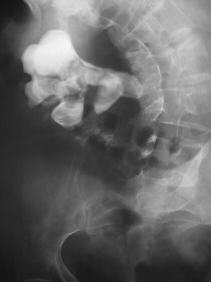
Staghorn calculus filling the renal collecting system (abdominal X-ray)
Nevit Dilmen / CC BY-SA 3.0 — Wikimedia Commons
Investigations
Non-contrast CT KUB (gold standard — stone, size, site, hydronephrosis); urine dip (haematuria, infection), MSU; U&Es (renal function), calcium/urate; stone analysis; ultrasound (pregnancy/children to avoid radiation).
Management
NSAID analgesia; <5 mm pass spontaneously / alpha-blocker; size/site-based removal (SWL/URS/PCNL)
- 1Confirm with CT KUB and give an NSAID for analgesia. Small stones (<5 mm) usually pass — watchful waiting ± an alpha-blocker (medical expulsive therapy) for distal ureteric stones <10 mm.Gate: An obstructed AND infected system (fever/systemic upset) → EMERGENCY decompression (nephrostomy/stent) + antibiotics; and never accept 'renal colic' in an older arteriopath without excluding a ruptured AAA.
- 2Active removal by size/site (NG118): ureteric 20 mm or staghorn → PCNL. Investigate/prevent recurrence (hydration, stone analysis, metabolic work-up).
Key points
Loin-to-groin colic + restless patient + haematuria = stone → CT KUB, NSAID analgesia. <5 mm pass; alpha-blocker for distal ureteric <10 mm; SWL/URS/PCNL by size. EMERGENCY = obstructed + infected (pyonephrosis) → decompress + antibiotics. And exclude a ruptured AAA in older patients.
Monitor & prognosis
Pain/stone passage, renal function, infection; recurrence prevention.
Most small stones pass; recurrence is common — prevention matters.
Source: NICE NG118 (renal & ureteric stones)