Pleural effusion
Fluid in the pleural space — transudate (systemic) vs exudate (local) by Light's criteria
Overview
Accumulation of fluid in the pleural space. The fundamental split is transudate (low protein — heart/liver/renal failure, hypoalbuminaemia) vs exudate (high protein — infection, malignancy, PE, inflammation), separated by Light's criteria. Diagnostic aspiration (with pleural fluid analysis) is the key step; a purulent/low-pH effusion is an empyema needing a drain.
Recognise
- Breathlessness, pleuritic pain; STONY DULL percussion, reduced breath sounds and reduced vocal resonance over the effusion; tracheal deviation away if large
- CXR: blunting of the costophrenic angle, meniscus; ultrasound guides aspiration and detects loculations
- Transudate (30 g/L): infection, malignancy, PE, inflammation
Red flags
- Empyema (purulent fluid, pH <7.2, low glucose) → chest drain (see empyema/abscess)
- Massive effusion with respiratory compromise → therapeutic drainage; haemothorax/malignant effusion → specific management
Differentials & how to tell them apart
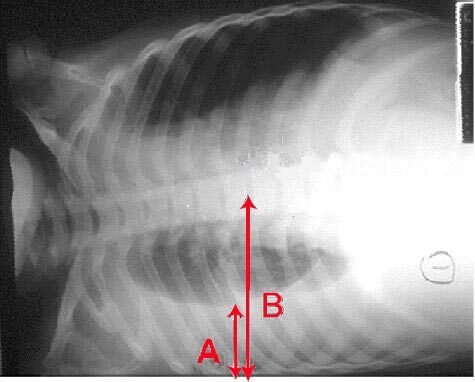
Pleural effusion — blunting of the costophrenic angle with a meniscus (chest X-ray)
InvictaHOG / Public domain — Wikimedia Commons
Investigations
CXR + ULTRASOUND; diagnostic pleural ASPIRATION — protein, LDH (Light's criteria), pH, glucose, cytology, microscopy/culture; LIGHT'S CRITERIA for borderline protein (exudate if fluid:serum protein >0.5, fluid:serum LDH >0.6, or fluid LDH >⅔ upper normal serum); CT/pleural biopsy for suspected malignancy.
Management
Aspirate + Light's criteria → treat the cause; drain symptomatic/empyema/malignant effusions
- 1Confirm clinically (stony dull base) and on CXR/ultrasound, then perform a diagnostic pleural aspiration. Apply Light's criteria to classify transudate vs exudate when protein is borderline.Gate: Transudates (heart/liver/renal failure) are treated by addressing the systemic cause — not routinely drained; a purulent/low-pH (<7.2) parapneumonic effusion is an empyema needing a chest drain.
- 2Exudates need the local cause found (malignancy → cytology/CT/biopsy; infection → drain); therapeutic/large effusions → controlled drainage; malignant effusions → pleurodesis or indwelling catheter.
Key points
Stony dull base + reduced breath sounds = pleural effusion → ultrasound-guided aspiration + LIGHT'S CRITERIA (transudate 30 g/L). Transudate (heart/liver/renal) → treat the cause, don't routinely drain; exudate → find the local cause; pus/pH <7.2 = empyema → chest drain.
Monitor & prognosis
Resolution/recurrence, the underlying cause; cytology/biopsy results for malignancy.
Depends on the cause; malignant effusions recur (pleurodesis/catheter help).
Source: BTS pleural disease