Iron-deficiency anaemia
Iron deficiency (blood loss, malabsorption, poor intake, increased demand) → microcytic hypochromic anaemia
Overview
The commonest anaemia worldwide — a microcytic, hypochromic anaemia from depleted iron stores. The cause matters more than the number: in adults (especially men and post-menopausal women) it signals occult GI blood loss / malignancy until proven otherwise; in menstruating women, menorrhagia; consider coeliac disease (malabsorption) and dietary deficiency. Treat with oral iron and find and treat the cause.
Recognise
- Fatigue, pallor, breathlessness; signs of chronic deficiency — koilonychia (spoon nails), angular stomatitis, glossitis, pica
- Microcytic hypochromic film (pencil cells, anisocytosis); LOW ferritin (the key test — but ferritin is an acute-phase reactant, can be falsely normal/high with inflammation)
- Causes: blood loss (GI — ulcer/cancer/angiodysplasia; menstrual), malabsorption (coeliac), poor intake, increased demand (pregnancy/growth)
Red flags
- Iron deficiency in any adult man or post-menopausal woman → urgent investigation for GI cancer (2-week-wait OGD + colonoscopy)
- Coeliac disease as a cause of malabsorption → test (anti-TTG)
Differentials & how to tell them apart
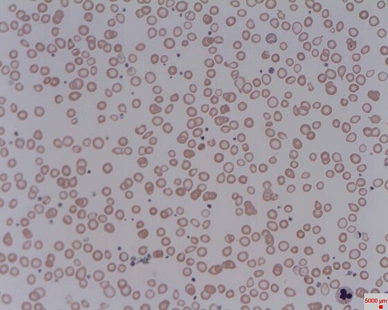
Iron-deficiency anaemia — hypochromic microcytic red cells (blood film)
Prof. Osaro Erhabor / CC BY-SA 3.0 — Wikimedia Commons
Investigations
FBC (microcytic hypochromic), ferritin (low — diagnostic; interpret with CRP as it rises with inflammation), iron studies (low iron, high transferrin/TIBC); blood film; coeliac serology (anti-TTG); upper + lower GI endoscopy in adults to find the bleeding source; urinalysis/menstrual history.
Management
Oral iron + find/treat the cause (GI source, menorrhagia, coeliac)
- 1Confirm with a low ferritin (microcytic hypochromic film) and start oral iron; alternate-day dosing aids absorption and continue for ~3 months after the Hb normalises.Gate: The cause is the priority: iron deficiency in any man or post-menopausal woman → urgent 2-week-wait OGD + colonoscopy for GI cancer; test for coeliac disease; ferritin can be falsely normal with inflammation (check CRP).
- 2IV iron if oral is not tolerated/absorbed or response is inadequate; transfuse only for symptomatic/severe anaemia; treat the underlying source.
Key points
Microcytic hypochromic anaemia + LOW ferritin = iron deficiency → oral iron (alternate-day, continue 3 months after Hb normal) AND find the cause: any man / post-menopausal woman → GI cancer work-up; women → menorrhagia; consider coeliac. Ferritin is an acute-phase reactant (falsely normal with inflammation).
Monitor & prognosis
Hb/reticulocyte response (recheck at 2–4 weeks), ferritin replenishment, the cause.
Excellent once iron is replaced and the source treated.
Source: NICE CKS / BSH iron deficiency